睾丸的正常发育及充分下降是雄性生殖与内分泌功能的重要保证,涉及诸多泌尿生殖系统疾病。既往对睾丸下降的研究大多是局部的或集中在睾丸下降以后,且常将睾丸作为单独的研究对象。睾丸下降是一个连续的、动态的过程,期间涉及睾丸形态、位置、功能改变,也伴随诸多周边毗连组织的明显变化,其中睾丸引带、附睾、腹股沟管等与之关联特别密切,但它们在发育过程中的相互作用一直未明,为此,本实验运用三维组织学重建(3Dhr)技术从显微形态学水平,对睾丸下降过程中睾丸引带、附睾、腹股沟管等的形态位置变化进行了纵向时序上的观察研究。现报道如下。
1 材料与方法1.1 标本准备昆明小鼠,胎鼠于母鼠怀孕(GD)第15、17、19天(GD15、GD17、GD19)行颈椎脱臼法处死后获取,仔鼠标本待母鼠自然分娩后第3、7天(PD3、PD7),经二氧化碳(CO2)窒息处死后获取。以上标本置于40 g/L多聚甲醛固定24 h后切取肾脏平面以下组织整块石蜡包埋。
1.2 连续切片制备及染色把包埋好的组织块置冷冻切片机内(-12 ℃)冷冻20~30 min,后按20 μm连续切片,每张切片在展片机水槽中(水温48 ℃)充分展开,捞片时分别按奇数偶数切片贴于同一序列载玻片上。实验时取基数或偶数序列片进行HE染色,其余切片备份保存。
1.3 计算机处理染色后的组织切片使用PerkinElmer全自动智能切片分析系统(美国)进行拍摄。每张切片均10×4倍拍摄成一张全景图片并保存。后运用Photoshop软件进行图片对齐,运用3D-doctor软件进行相关组织、器官边界勾勒及三维重建,调节X、Y、Z各轴参数,获得需要及满意的三维模式图。
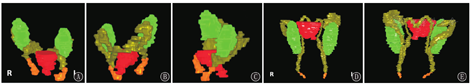
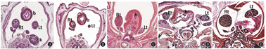
2 结果2.1 小鼠睾丸、附睾及睾丸引带三者位置关系睾丸、附睾及睾丸引带三者位置关系密切(图1)。3Dhr模式图显示:附睾与睾丸紧密相邻,并将睾丸包绕其中;附睾尾部与睾丸引带相连接,睾丸与引带无直接连接关系(图2A、图2B)。睾丸在孕期时其上极高出附睾头部,下极与附睾尾部、体部及输精管紧密连接,出生后附睾头部、体部及尾部则将其如"C"形包绕,睾丸在整个下降过程中形态变化小,始终如长椭圆状(图1A、图1D)。睾丸引带在孕期时体积相对较大,形如椎体状(图1A,图2C),出生后退化变得细小如条索状(图1D,图2D),孕期及生后睾丸引带的体积均明显小于睾丸;附睾在孕期时形态很不规则,附睾尾部及输精管堆积体积相对较大,其横径与睾丸横径相当(图1A,图2E),生后随着睾丸降入阴囊,附睾体、尾部及输精管变得细长,附睾尾部始终在睾丸以下(图1D、图1E,图2F)。
点击查看大图
图1
小鼠睾丸、附睾、引带、膀胱(颈)三维组织学重建模式图 A、B、C分别为怀孕第17天正面观、背面观、侧面观,见附睾体、尾与输精管堆积体积较大,分界不清;D、E分别为出生第3天正面观、背面观,见附睾体、尾及输精管拉长变细,体积变小,分界清晰
Figure 1
Three dimensional histology reconstruction models of testicle,epididymis,banding and bladder (neck) in mice A,B and C are frontal,dorsal and lateral views at gestation day 17,respectively.These models show that the epididymis,tail and vas deferens accumulation volume is large,and the boundary is not clear.D and E are respectively frontal and dorsal views at postnatal day 3.The epididymis,tail and vas deferens are elongated and thinner,the volume is smaller,and the boundary is clear
点击查看大图
图2
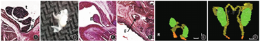
睾丸(t)、附睾(e)、引带(g)三维组织学重建模式图及对应二维组织平面及大体图 A、C分别为GD19睾丸引带平面纵切、横切面(×400);B:PD3睾丸、附睾、引带及输精管(sd)活体组织;D:PD3引带平面横切面,显示附睾包绕睾丸,睾丸引带连接于附睾尾部,随时间引带退化体积变小(×400);E:GD19;F:PD3
Figure 2
Three dimensional histology reconstruction models and the corresponding two-dimensional picture of the testicle (t),epididymis (e) and gubernaculums (g) A and C are longitudinal and transverse sections of the testicular ejection zone at GD19,respectively(×400);B:testis,epididymis,gubernaculum,and spermatic duct (sd) zonal living tissue at PD3;D:Cross section of PD3 lead strip plane.The epididymis is shown to wrap around the testicles.The gubernaculum is attached to the tail of the epididymis(×400);E:GD19;F:PD3
点击查看大图
图2
睾丸(t)、附睾(e)、引带(g)三维组织学重建模式图及对应二维组织平面及大体图 A、C分别为GD19睾丸引带平面纵切、横切面(×400);B:PD3睾丸、附睾、引带及输精管(sd)活体组织;D:PD3引带平面横切面,显示附睾包绕睾丸,睾丸引带连接于附睾尾部,随时间引带退化体积变小(×400);E:GD19;F:PD3
Figure 2
Three dimensional histology reconstruction models and the corresponding two-dimensional picture of the testicle (t),epididymis (e) and gubernaculums (g) A and C are longitudinal and transverse sections of the testicular ejection zone at GD19,respectively(×400);B:testis,epididymis,gubernaculum,and spermatic duct (sd) zonal living tissue at PD3;D:Cross section of PD3 lead strip plane.The epididymis is shown to wrap around the testicles.The gubernaculum is attached to the tail of the epididymis(×400);E:GD19;F:PD3
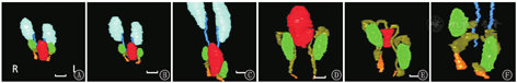
2.2 小鼠睾丸下降过程动态观察GD15时,双侧睾丸位于肾下极,准备开始下降;GD17时,双侧睾丸下降至膀胱中部位置,开始远离肾脏;GD19时,右侧睾丸下降速度减缓,仍位于膀胱中部位置,而左侧睾丸则下降至膀胱下部位置,此时双侧睾丸明显远离肾脏;PD3时,右侧睾丸下降速度加快,此时双侧睾丸位置大致相当,均位于膀胱下部位置;PD7时,左侧睾丸加速下降,降于膀胱颈部以下,而右侧睾丸下降缓慢,位于膀胱颈部附近。小鼠睾丸在PD7以后下降完全,最终是双侧睾丸位置不完全对称,左侧睾丸稍低于右侧(图3,图4,图5)。
点击查看大图
图3
小鼠睾丸下降过程三维组织学重建模式图(正面观) A:GD15;B:GD17;C:GD19;D:PD3;E:PD7;F:C移除膀胱后局部放大。显示GD15时双侧睾丸位于肾下极平面附近,GD17时睾丸降至膀胱中部以上,GD19时左侧睾丸降至膀胱中部以下,右侧睾丸仍在膀胱中部以上;PD3时双侧睾丸基本位于同一水平,降至膀胱中部以下,到PD7时左侧睾丸位置(最终)低于右侧
Figure 3
Three dimensional histology reconstruction of testicular descent in mice (frontal view) A:GD15;B:GD17;C:GD19;D:PD3;E:PD7;F:C local magnification after bladder removal.The models showed that at GD15,both testicles were located near the plane of the lower pole of the kidney;at GD17,the testicles fell above the middle of the bladder;at GD19,the left testicle fell below the middle of the bladder;and the right testicle remained above the middle of the bladder.At PD3,both testicles were basically at the same level and fell below the middle of the bladder.At PD7,the left testicle has the final position lower than that of right
点击查看大图
图4
小鼠睾丸下降过程三维组织学重建模式图 A:GD15;B:GD17;C:GD19为侧面观;D:PD3;E:PD7为背面观;F为C移除膀胱后局部放大并旋转图像
Figure 4
Three dimensional histology reconstruction model of testicular descent in mice A:GD15;B:GD17;C:GD19 (lateral view);D:PD3;E:PD7 (dorsal view);F is C enlarged and rotated,and with the bladder removed
点击查看大图
图4
小鼠睾丸下降过程三维组织学重建模式图 A:GD15;B:GD17;C:GD19为侧面观;D:PD3;E:PD7为背面观;F为C移除膀胱后局部放大并旋转图像
Figure 4
Three dimensional histology reconstruction model of testicular descent in mice A:GD15;B:GD17;C:GD19 (lateral view);D:PD3;E:PD7 (dorsal view);F is C enlarged and rotated,and with the bladder removed
点击查看大图
图5
小鼠睾丸下降平面图 A:GD15,左侧睾丸(Lt)出现时平肾脏(k)平面;B:GD17,左侧睾丸出现时肾平面已消失,大致到达膀胱(b)上部平面;C:GD19,左侧睾丸出现时大致平膀胱下部平面;D:PD3,左侧睾丸与右侧睾丸(Rt)大致同时出现,到达膀胱下部平面;E:PD7,左侧睾丸出现时膀胱消失,到达尿道(u)平面(×400)
Figure 5
Two-dimensional pictures of testicular descent in mice A:GD15,the left testicle (Lt) appeared flat in the renal (k) plane;B:GD17,when the left testicle appeared and roughly reached the upper bladder (b) plane,and the renal plane had disappeared;C:GD19,the left testicle was roughly flat on the lower surface of the bladder;D:PD3,the left and right testicles (Rt) appeared at roughly the same time,reaching the lower surface of the bladder;E:PD7,upon the appeara-nce of the left testicle,the bladder disappeared and reached the urethral (u) plane (×400)
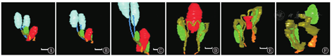
2.3 小鼠附睾形态衍变及下降过程3Dhr模式图显示:小鼠附睾在整个下降过程中形态变化大,先启动下降,并先于睾丸降至阴囊,附睾尾部始终位于睾丸下方(图6A~C)。在GD15,睾丸开始下降,此时睾丸上极高于附睾头,附睾体部及尾部相互堆积,位于睾丸下极以下,界限不清,体积较大(图6A);GD19时,睾丸准备进入阴囊,此时附睾体部及尾部仍堆积一起,体积很大,其横径大于睾丸横径(图6B);出生后PD3,睾丸降入阴囊,此时附睾体部、尾部及输精管拉长变小,界限较清,附睾将睾丸包绕其中,附睾尾部位于睾丸下极以下(图6C、图6D)。
点击查看大图
图6
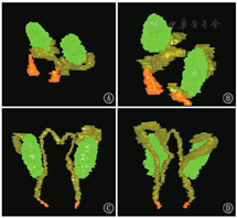
小鼠附睾形态衍变三维组织学重建模式图 A:GD15;B:GD19侧面观;C:PD3正面观;D:PD3背面观(各图经适当放大仅示意附睾形态,不代表真实比例)
Figure 6
Three dimensional histology reconstruction models of the morphological evolution of the epididymis in mice A:GD15;B:GD19 side view;C:PD3 frontal view;D:PD3 dorsal view (the figures are appropriately enlarge to show the shape of epididymis,which does not represent the true proportion)
点击查看大图
图6
小鼠附睾形态衍变三维组织学重建模式图 A:GD15;B:GD19侧面观;C:PD3正面观;D:PD3背面观(各图经适当放大仅示意附睾形态,不代表真实比例)
Figure 6
Three dimensional histology reconstruction models of the morphological evolution of the epididymis in mice A:GD15;B:GD19 side view;C:PD3 frontal view;D:PD3 dorsal view (the figures are appropriately enlarge to show the shape of epididymis,which does not represent the true proportion)
3 讨论睾丸是泌尿生殖系统的核心器官,胚胎期其正常发育与正常下降过程相辅相成,对睾丸下降过程的准确了解不仅有利于获知正常的生殖泌尿系统发育机制,更有利于明了该系统疾病及畸形的发育学病因,以便更精确地指导临床诊疗[1,2]。
睾丸下降是一个连续的、动态的过程,它不仅是睾丸形态、位置的改变,也同时伴随毗连组织结构的系列变化,如头侧的悬韧带、尾侧的引带、周围的腹股沟管结构及直到出生后并伴随终生的附睾、鞘膜等[3]。既往对睾丸下降的研究比较集中在调控机制方面,如各种因子、激素[4,5,6]。而对睾丸毗邻结构的研究较少,有的也多是局部的和某个特定时间点的。这与睾丸下降发育涉及多种组织器官,机制及过程相当复杂,期间(胚胎发育期)各结构形态、位置随时间变化、移位巨大(有的甚至消失)等相关,它们给各种研究带来很大困难[7,8]。
随着3Dhr技术的发展,为以小动物为模型,在显微水平系统地、精细甚至精确地时序性动态观察研究睾丸及其关联结构的同步变化提供了可能。3Dhr技术基于已广为应用的CT、磁共振成像(MRI)及B超等影像3D重建技术的思路,由于其二维图像信息取自生物显微图像(而非影像),其分辨率无可比拟,但同时,由于图像的旋转配准等没有影像设备及其配套软件的"自动化"帮助,3Dhr的许多技术难题还在不断克服中。目前,在神经、肌肉等通常难于认识的领域,3Dhr已有不少成功的研究应用[9,10]。提示其不仅可用于研究组织器官的整体或局部,还可进行细胞、细胞器水平的重建,甚至借助特殊染色深入到分子水平研究[11],使微观范围内的空间构象得到展现。因此,3Dhr对于器官组织的精细(显微水平)形态学研究很有潜力。
本课题组也一直在探索3Dhr技术应用于显微组织结构及雄性小鼠泌尿生殖系统研究的可行性[12,13]。在本实验中,观察到睾丸上极与附睾头部连接,向下逐步通过疏松结缔组织与附睾体、尾部连接,睾丸引带为睾丸下极与附睾尾部周围的组织向下延续而成,它锚定附睾尾部及睾丸下极(附睾头及睾丸上极由精索锚定)。这些发现多与所认识的大体解剖结构一致,但也更清楚地表明睾丸引带不是直接锚定于睾丸本身。此外,根据时序性3Dhr图像观察,发现睾丸左右侧和同侧不同时段其下降速度是不一样的,开始左侧睾丸下降快,睾丸下降第二阶段启动后,右侧下降加快,后左侧再加快,并在最终位置低于右侧。
既往研究认为,睾丸下降开始时,头侧的悬韧带发生退化,尾侧的引带则发生"膨大反应",体积不断增大,长度相对减小,从而使引带有一定张力,牵引睾丸下降至内环口处[3]。此外,引带发生膨大,亦会将内环口扩张,为睾丸进入腹股沟管做准备。本研究发现,胚胎期睾丸引带确实发生膨大,但其体积明显小于睾丸体积,扩张内环口后不足以使睾丸通过,而睾丸引带连接附睾尾部,首先牵引的是附睾尾部下降到内环口。在孕期,附睾体、尾部及输精管堆积成团,体积较大,而睾丸在整个下降过程中的形态并无较大改变。因此,提示睾丸引带膨大扩张内环口后,牵引附睾尾进入腹股沟管,附睾尾部进入后体部及输精管亦相继进入,它们通过形态的改变不断扩张内环口及腹股沟管,使其空间足够大以利睾丸顺利通过。这可能是更合理的睾丸下降过程。
在睾丸进入阴囊阶段,过去研究认为,膨大的睾丸引带同时扩张阴囊并引导睾丸通过扩张的腹股沟管进入阴囊,同时引带逐步缩短退化,含水量不断减少,纤维成分不断增加,最终退化成纤维带,锚定睾丸于阴囊内,大体解剖上基本消失[14]。本研究观察到,睾丸引带在小鼠出生后(相当于人睾丸经腹股沟阴囊下降阶段)即开始退化、缩短,体积相对不断变小,引导附睾尾部及睾丸下极逐步通过腹股沟管降入阴囊。整个过程附睾尾部均在睾丸下极下方,睾丸引带锚定在它们周围组织并固定于阴囊内或附近。整个过程附睾尾部、体部及头部成"C"形将睾丸上下包绕。
过去,在睾丸下降过程的研究中,对附睾的研究和描述甚少。本研究证实,附睾在睾丸下降过程中其形态、位置也发生巨大变化,加之其形态不规则,二维研究确实很难准确、合理地描述。睾丸下降始终伴随着附睾下降,附睾尾部始终位于睾丸以下,附睾下降睾丸才随之下降[15]。也有研究提到附睾下降机制可能就是睾丸下降机制[16]。在临床上,隐睾患儿中有相当部分伴附睾畸形[2,8],后者与生育相关。但附睾与睾丸形态、位置变化(下降)的关系还研究得很不足。本研究观察发现,附睾下降应该是睾丸下降的初始触发点,睾丸引带首先牵引的是附睾尾部下降,附睾尾部在整个下降过程及下降完成后始终位于睾丸下极之下,通过观察它们的形态改变及位置变化可以比较客观地阐述这种过程。或许,从附睾作为出发点进行睾丸下降的研究是值得重视的,这对于睾丸下降过程中发生的异常或许有新的指导意义。不过这种设想仍需进一步研究以求更加完善、准确。
综上,以小鼠模型结合组织块连续显微切面及3Dhr技术能有效的、时序性系统地研究观察睾丸及其毗邻组织器官的立体形态及变化。并据此推测睾丸下降过程应是:睾丸引带起引导作用,确保附睾、睾丸下降沿正确的方向;睾丸引带牵引附睾尾部(及睾丸)下降至内环口,附睾尾部及体部通过其形态改变不断扩张内环口及腹股沟管,使它们足够大而适合睾丸通过;附睾先进入阴囊后继续扩大阴囊,为睾丸进入提供足够空间,然后睾丸顺着附睾(体)下降直至阴囊。


